I don’t understand the image examination report, I don’t know which department to look for to interpret it after taking a bunch of films, and the examination conclusion shows "nothing unusual". Do you want to ask the doctor again … For the patients’ doubts, Lin Qing (pseudonym), a 42-year-old chest disease patient, easily solved them in the interpretation clinic of shanghai changzheng hospital Radiodiagnosis Department.
"What are the specific changes in the disease, whether it is serious or not, and how long it takes to review it, the doctors here will answer it professionally and patiently for the first time." To Lin Qing’s great praise, there is no sense of tension and oppression in seeing a doctor here. In his words, when getting along with doctors, he completely forgot his identity as a patient.
"Our goal is to cultivate a doctor with temperature." According to Professor Liu Shiyuan, director of the Department of Radiological Diagnosis in shanghai changzheng hospital, only by "moving" medical images to clinics and wards can we truly realize the transformation from "image" as the center to "patient" as the center.
In fact, Professor Liu Shiyuan, who has just taken over as the chairman of the 16th Committee of Radiology Branch of Chinese Medical Association, also regards medical image AI as one of the "magic weapons" to improve medical service actions and enhance people’s medical experience. Early screening and early diagnosis of chest diseases with AI is a typical example. At the same time, as the chairman of the Asian Chest Radiology Society, the vice chairman of the Radiologist Branch of the Chinese Medical Doctor Association, and the chairman of the China Medical Imaging AI Industry-University-Research Innovation Alliance, Professor Liu Shiyuan has a keen sense of smell and unique views on the development of medical imaging.
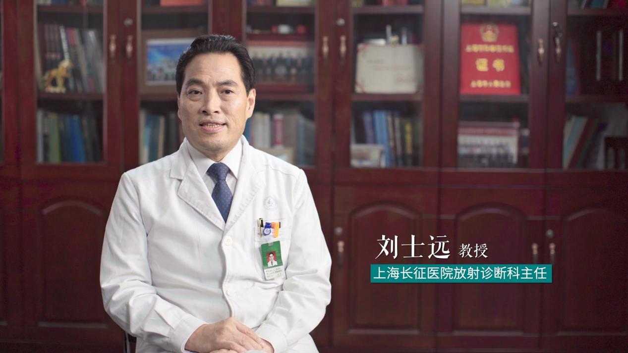 Professor Liu Shiyuan, Chairman of the 16th Committee of Radiology Branch of Chinese Medical Association and Director of Radiological Diagnosis Department of shanghai changzheng hospital.
Focus on the "five modernizations"
The so-called "a glimpse of the whole leopard."
As an important basis for disease diagnosis and treatment, medical imaging has become an indispensable part of doctors’ disease diagnosis and treatment. "We have never been so irreplaceable." Professor Liu Shiyuan introduced that at present, 70% of clinical diagnostic information comes from medical images. With the progress and development of medicine, computer, big data, artificial intelligence, internet and other technologies have promoted the revolution and innovation of imaging equipment, making the role of imaging department more and more powerful and multidimensional.
"In the future, China medical imaging will develop in the direction of’ five modernizations’ under the premise of taking patients as the center, emerging technologies as the carrier and all-round systems as the guarantee." The "five modernizations" referred to by Professor Liu Shiyuan are precision, intelligence, clinic, prehospital and networking;
1. Precision means upgrading the image business, realizing precision medicine, individualizing diagnosis and treatment, maximizing the effect and minimizing the side effects.
2. Intelligentization is the cross-integration of "doctor +AI" to maximize the diagnostic efficiency, improve the work efficiency of doctors, and change the working mode and process.
3. Clinicalization means that the imaging department is transformed from the traditional auxiliary diagnosis department and medical technology department into a clinical discipline, and goes deep into outpatient clinics and wards; From "computer-centered" to "patient-centered".
4. Pre-hospital refers to taking disease prevention as the center, going out of the hospital and changing medical imaging from passive service to active service; From "disease-centered" to "health-centered".
5. Networking is based on the Internet to break through the time and space constraints, realize the integration of in-hospital and out-of-hospital services, form a patient-centered global coverage medical imaging service system, and help the sinking of high-quality medical resources and grassroots improvement.
As the leader of the discipline, Professor Liu Shiyuan has led the Department of Radiological Diagnosis of shanghai changzheng hospital to set sail on the new course of benchmarking.
Strip combination open circuit
Nowadays, medical imaging plays an important role in the whole process of clinical diagnosis and treatment, such as quantitative, qualitative, staging, prognosis prediction, treatment choice, discharge follow-up, interventional therapy, hybrid operating room, etc., all of which require the guidance and participation of medical imaging, and the traditional medical imaging model can no longer meet the needs of clinical diagnosis.
How to transform the imaging department from the traditional auxiliary diagnosis department and medical technology department into a clinical discipline to better serve the clinic and patients? This is the "homework" that Professor Liu Shiyuan pays more attention to in his heart.
In the early 1990s, the Long March Hospital was in line with international standards and took the lead in implementing the integrated management mode of imaging discipline in China.
"The so-called’ compartmentalization’ means that departments are divided into X-ray, CT, MRI, nuclear medicine and other departments according to the examination module to complete patient examination; Diagnostic doctors are divided into neurology group, head and neck group, osteology group, cardiothoracic group, abdominal group, etc. Doctors in each group can see images of X-ray, CT, MRI, nuclear medicine and other imaging equipment at the same time, and are committed to creating the concept of’ big image’. " In Professor Liu Shiyuan’s view, the subdivision of sub-majors enables medical images to serve the clinic more carefully and professionally, and maximizes the effect of disease diagnosis, which is conducive to improving the quality of medical services and effectively promoting the output of scientific research results.
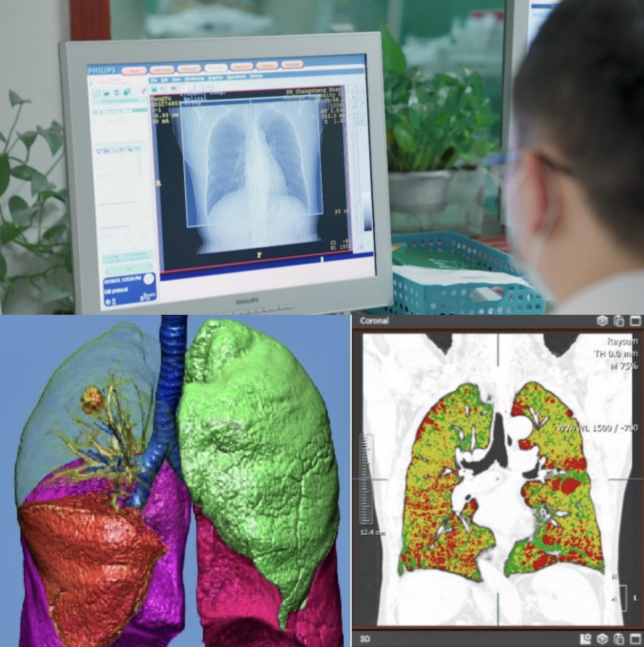 One-stop chest CT scanning can quantitatively and intuitively evaluate pulmonary nodules and COPD.
Sub-professional division of labor is also helpful for imaging doctors to deepen their understanding of clinical, scientific research and teaching problems of systemic diseases, and to form the characteristics of hospital imaging department and department doctors.
Director Liu Shiyuan has been engaged in imaging diagnosis of chest diseases for more than 30 years, which has not only formed a department and its own characteristics, but also formed a leading advantage in the country and a high reputation in Asia and the world.
Among the top 10 causes of death in the world announced by the World Health Organization in 2018, ischemic heart disease ranks first, chronic obstructive pulmonary disease ranks third and lung cancer ranks sixth. The incidence of these three chronic diseases in China ranks first in the world, with a total population of more than 150 million, resulting in a disease burden accounting for more than 70% of the total disease burden.
"Image is the key to improve the prevention and control level of major chronic diseases in the chest!" Professor Liu Shiyuan, who has achieved a lot in early screening of chest diseases, hit the nail on the head.
As the earliest department to start multi-center screening of major chest diseases, shanghai changzheng hospital Radiological Diagnosis Department has made great efforts to improve the early screening and early diagnosis of chest diseases: on the one hand, it has strengthened the configuration of hardware facilities, and the department currently has a complete set of imaging diagnostic equipment such as intelligent AI 256-slice CT, nuclear magnetic resonance, PET-CT, etc., and its overall hardware strength is at the advanced level at home and abroad; On the other hand, they use low-dose CT one-stop scanning technology to screen high-risk groups, and evaluate whether there are pulmonary nodules and malignant possibility, whether there is calcification in cardiovascular system and the possibility of sudden heart events, whether there is chronic obstructive pulmonary disease and early warning. "This low-cost way for one-stop evaluation of the three major chronic diseases in the chest can help to move forward the prevention and control of major chest diseases, improve the early diagnosis rate and reduce the mortality rate." Professor Liu Shiyuan said.
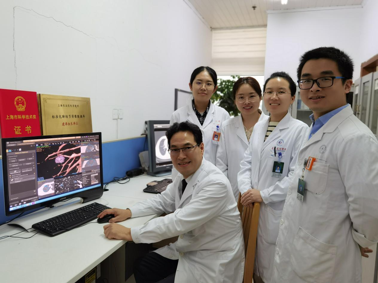 Professor Liu Shiyuan and the students
"In the long-term clinical and scientific research practice, we have gradually formed the characteristics of imaging diagnosis and interventional treatment of chest diseases. The overall diagnostic accuracy rate of lung cancer is 98.2%, and the diagnostic accuracy rate of early lung cancer is 95%." Professor Xiao Yi, deputy director of the Department of Radiological Diagnosis, introduced that in 2019, their research on key issues of early screening and early diagnosis of lung cancer based on multimodal imaging won the first prize of Shanghai Science and Technology Progress Award.
We should know that in clinic, judging the nature of pulmonary nodules depends on the location, size, shape and calcification of pulmonary nodules in CT scanning, but the morphological characteristics presented by conventional CT scanning are often difficult to meet the clinical needs. For this pain point, shanghai changzheng hospital Department of Radiology has its own "secret": ultra-high resolution CT target scanning and CT target reconstruction are very important in early lung cancer screening. They adopt the ultra-high definition CT scanning mode of 1024X1024 matrix, which can fully and clearly show the "details" of pulmonary nodules and make correct image diagnosis better. "Compared with high-definition images, blurred images are like a person walking into the prairie of clear Wan Li in a foggy environment, and there is a feeling of brightening up at the moment." Professor Xiao Yi described it.
It is worth mentioning that in the past, when using traditional CT for coronary artery examination, the scanning process of patients was complicated, heart rate control and respiratory training were indispensable, and the success rate and accuracy of scanning were not high. With the upgrading of CT equipment, the above problems have been optimized in the Department of Radiological Diagnosis in shanghai changzheng hospital.
The health community has learned that Philips Smart AI 256-slice CT screening for patients can not only effectively cope with all kinds of high heart rates, but also freeze coronary arteries through B2B technology, and automatically avoid abnormal heart rhythms through the unique ARAD smart heart engine to ensure the success of scanning to a greater extent.
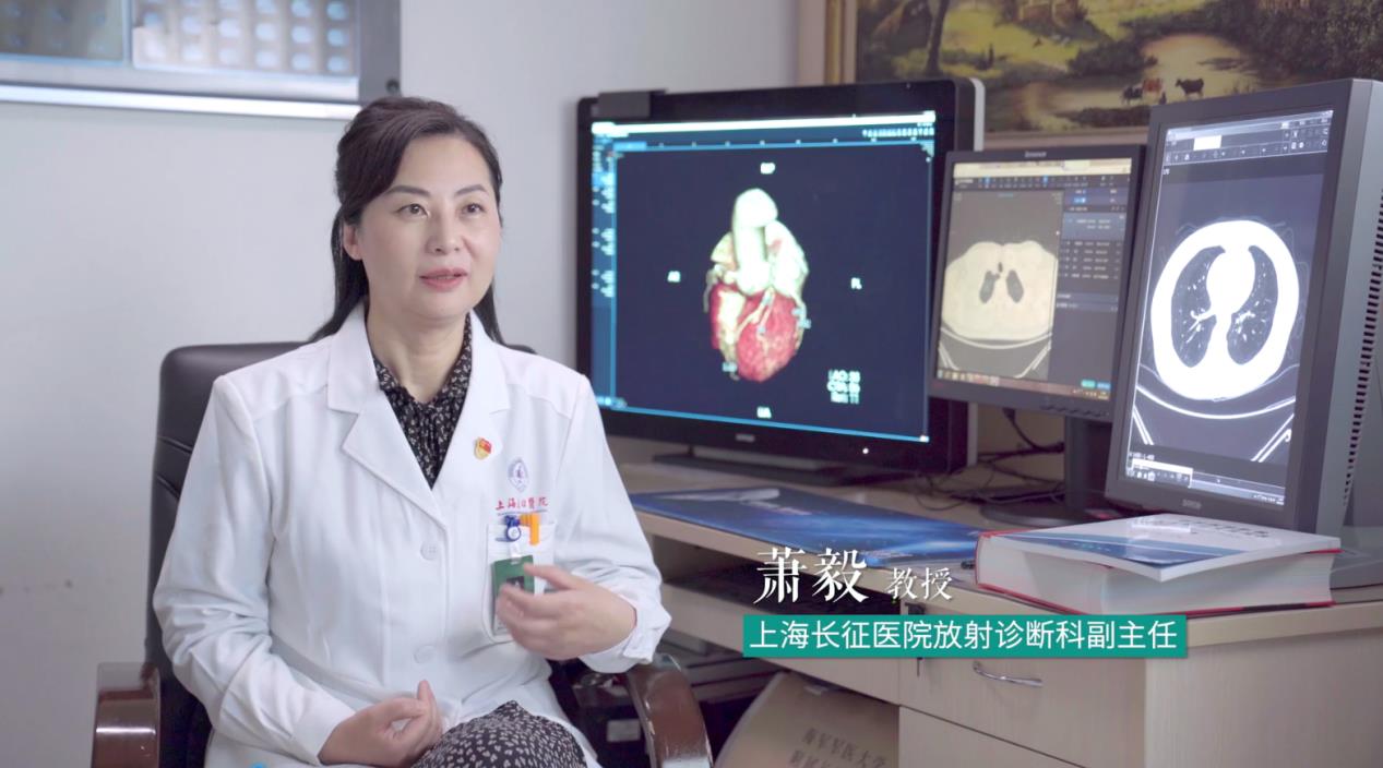 Professor Xiao Yi, Deputy Director of Radiological Diagnosis Department of shanghai changzheng hospital
In order to improve people’s awareness rate of chest diseases and screening cooperation, Professor Liu Shiyuan also took the lead in setting up a public welfare training program of "Cardiopulmonary Escort". "With the scientific research experience accumulated by our department for many years, the data can be deeply mined and analyzed. These three diseases can be evaluated by one-stop CT scanning, which has very high social value and reduces the medical burden of patients. Our previous cooperative research foundation with Royal Dutch Academy of Sciences on screening three major chest diseases is in line with the demand orientation of the National Healthy China 2030 Plan. Combining the team’s own advantages and the current integration of artificial intelligence, we can carry out more in-depth research, which will be a long-term research direction in the future. At the same time, we are also organizing the writing of a 100-question handbook on lung nodules, a handbook on major chronic diseases in the chest, various precautions for imaging examination, and the identification and disposal of contrast agent allergies to educate the public on science. " Professor Li Fan, Deputy Director, introduced.
In 2021, the Ministry of Industry and Information Technology and the National Health and Wellness Commission jointly issued the List of 5G+ Medical and Health Application Pilot Projects, and shanghai changzheng hospital was on the list. "This is a good opportunity." Professor Liu Shiyuan revealed his plan to the health sector. "Based on the Internet and 5G technology, we can promote and apply the low-dose CT one-stop early screening and early diagnosis technology for major chest diseases at the grassroots level, improve the detection rate of chest diseases at the grassroots level, and truly benefit the people."
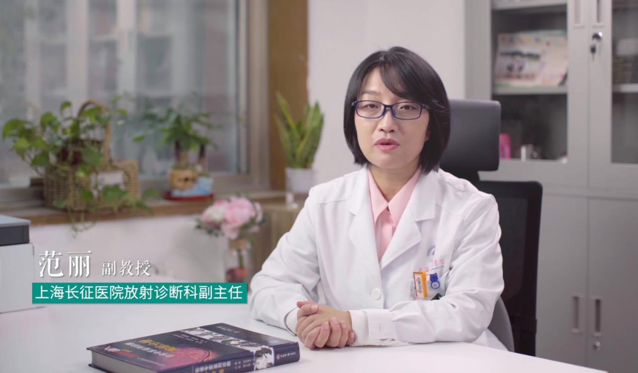 Professor Li Fan, Deputy Director of Radiological Diagnosis Department of shanghai changzheng hospital.
Wisdom image empowerment
With the change of disease spectrum, the difficulty of disease diagnosis increases, and the qualitative and quantitative requirements and clinical visualization process increase, which puts forward new requirements for image diagnosis.
Professor Liu Shiyuan introduced that at present, medical imaging faces the "three highs" dilemma of high work requirements, high work intensity and high work quality. According to the data of "China Artificial Intelligence+Medical Imaging Industry Market Research and Investment Prospect Forecast Report from 2018 to 2024", the annual growth rate of medical imaging data is 40%, while the annual growth rate of radiologists is only 4.1%. Front-line doctors need to write nearly 200 imaging reports a day, and there is a risk of missed diagnosis and misdiagnosis.
From 2015 to 2019, the state issued a series of policies to emphasize and encourage the application of artificial intelligence technology in the medical and health field, and smart medical care has gradually developed into a national strategy. As one of the earliest radiologists who embraced AI technology in China, Professor Liu Shiyuan told the health community that AI has four main advantages in medical imaging: disease detection, medical multidimensional measurement, accurate diagnosis, preoperative design and curative effect evaluation, and has covered lung, cardiovascular, bone and joint and other diseases.
What impresses Guan Yu, a senior doctor, is that the introduction of AI liberates imaging doctors from the front part of the screen and returns to the essence of doctors, at the same time, it improves the efficiency of imaging work and optimizes the workflow.
"Images are the eyes of clinicians, and the emergence of artificial intelligence has given our imaging doctors more power and more time, enabling us to better serve patients." Professor Xiao Yi takes coronary CTA as an example to directly attack the clinical pain points of imaging doctors. "If coronary CTA wants to obtain high-definition three-dimensional images, it can only be reconstructed in the post-processing workstation of hospital imaging department, which requires a lot of time and energy."
However, the combination of coronary CTA and AI can quickly complete the work of lesion identification, quantitative measurement and report output after the patient completes the heart scan, which greatly improves the inspection efficiency. "My post-processing time has been shortened from 30 minutes to 1 minute now. It is equivalent to the moment when the patient leaves the scanning bed, and I can see the reconstructed image in the office. " Professor Xiao Yi praised.
At the same time, they also make full use of the image diagnosis platform covering many clinical fields of radiology, such as heart and lung, to realize multi-class image fusion, and provide doctors with advanced visualization processing of multi-modal images, disease image feature mining and longitudinal tracking of lesions. "The progress of imaging technology provides reference for accurate, non-invasive and rapid clinical analysis, further improves the accuracy of diagnosis, and also reflects the humanistic care for patients." Li Qingchu, a radiation diagnosis technician, said.
"The preoperative 3D visual assessment of cardiovascular diseases allows doctors to clearly and intuitively see the three-dimensional disease structure on the computer, which plays a very good auxiliary role in clinical surgical treatment." Professor Xiao Yi introduced that it is one of the improvements of their medical service model to go deep into the clinical front line and participate in clinical diagnosis and treatment in an all-round way and formulate intuitive image reports according to clinical needs.
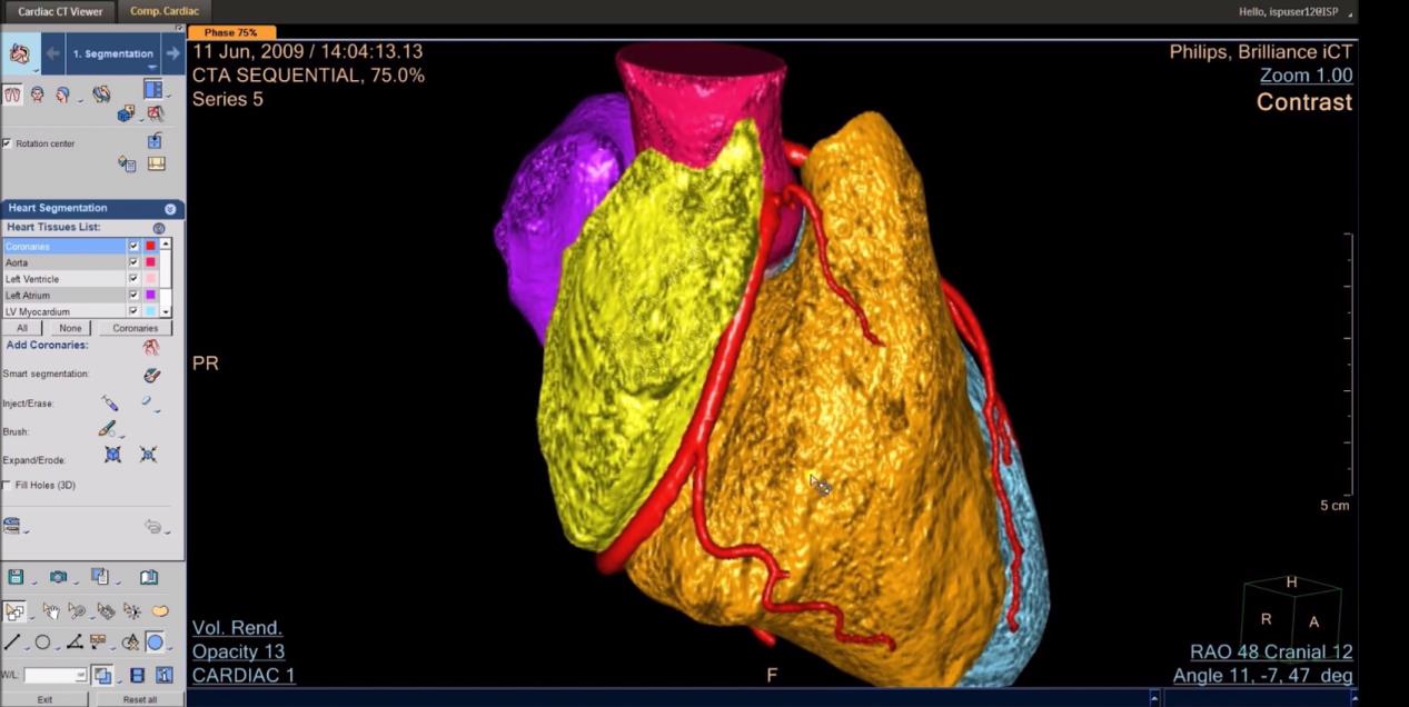 Three-dimensional image post-processing platform makes the disease characteristics more visual and three-dimensional, and can assist doctors to make rapid and accurate clinical diagnosis decisions.
Nowadays, medical imaging AI is gradually approaching some ideal clinical scenes of doctors, and has become a powerful "assistant" in everyone’s work. "Since 2017, the click-through rate of pulmonary nodule AI model has reached about 60%. From 2020 to now, the click-through rate has basically remained above 80%, and the clinical utilization rate has improved significantly." In this regard, Professor Liu Shiyuan is very pleased.
In order to promote the use of China medical imaging AI in Industry-University-Research, Professor Liu Shiyuan led the establishment of China Medical Imaging AI Industry-University-Research Innovation Alliance, and started the construction of standardized image database in China. Authoritative imaging experts in the United Nations wrote and published the China White Paper on Medical Imaging AI and the Report on the Development of Medical Imaging Artificial Intelligence 2020.
"Medical imaging AI is showing a variety of good development trends: First, the variety of products is becoming more and more abundant; Second, the product function is vertical and deepened; Third, the development of single disease to multi-disease and multi-task model; The fourth is the integration of software and hardware; The fifth is to cover the whole clinical process; Sixth, it is becoming more and more platformized; Seventh, based on new infrastructure, accelerate the integrated development of online and offline. " Professor Liu Shiyuan judged.
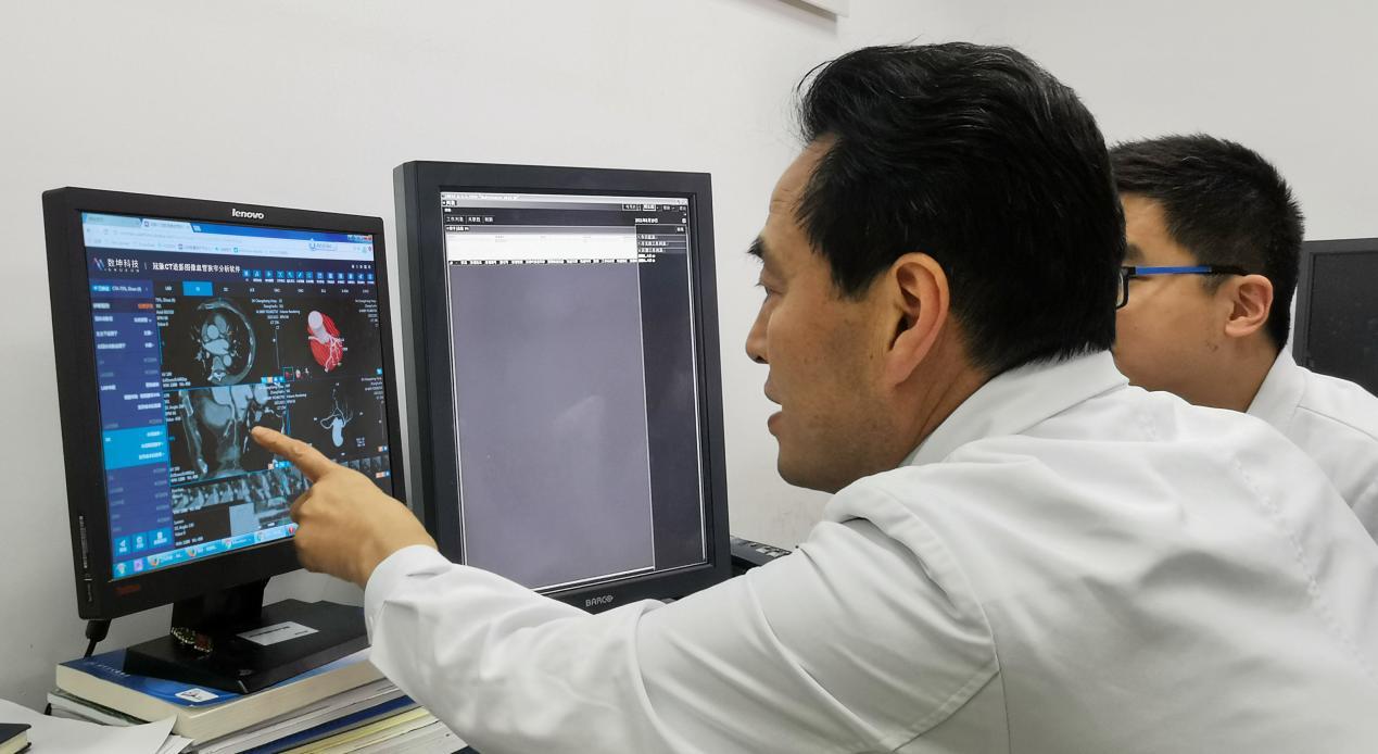 Professor Liu Shiyuan’s daily teaching
Talent construction ascends.
What kind of diagnostic thinking should a qualified imaging doctor have?
Professor Liu Shiyuan summed up his successful experience as a 16-word formula: comprehensive observation, careful analysis, clinical combination and diagnosis.
Professor Liu Shiyuan remembered a case 20 years ago. At that time, a patient diagnosed with advanced tumor by a foreign hospital came to see Professor Liu Shiyuan for consultation, but Professor Liu Shiyuan found that the patient’s diagnosis report was abnormal. "We re-examined the patient through thin-layer scanning and found that it was not an advanced tumor, but an old focus of tuberculosis. The patient was very excited to hear the news." This also makes Professor Liu Shiyuan deeply realize that compound medical talents in imaging department are particularly important.
Professor Liu Shiyuan adopted a two-pronged approach to build a "sophisticated" talent echelon:
First, positively guide and stimulate the internal drive and creativity of department doctors.
"We have traditional teaching, such as expert series lectures; There are also English case reports, difficult case reading sessions, and flipping the classroom, so that students can listen to the teacher, and then everyone can ask questions and discuss them together to ignite the students’ thinking ability. " Professor Xiao Yi introduced to the health sector that they also used 3D printing technology to restore the diseased parts of patients in a ratio of 1:1, so that young doctors could improve their diagnostic skills through model exercises.
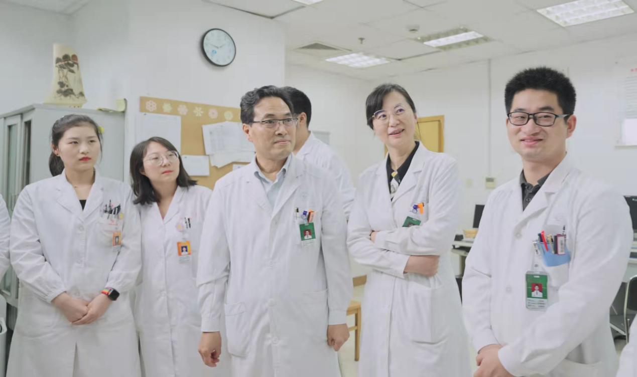 Professor Liu Shiyuan’s daily teaching
Second, please come in and send out. For example, send young doctors to study abroad, invite experts to share clinical experience in departments, and improve everyone’s professional ability.
Wang Xiang, a young doctor in the Department of Radiological Diagnosis in shanghai changzheng hospital, told the health sector, "Through intensive study in the past five years, I have initially cultivated good clinical diagnostic thinking, and my diagnostic level has been continuously improved, and I have published many SCI papers."
Moreover, as a national training base for imaging medical residents and a national continuing education base for radiologists, the Department of Radiology and Diagnostics of shanghai changzheng hospital has improved the primary medical ability through diversified teaching modes. Mou Yong, a doctor studying in Qingdao who has gained a lot, told the health community that "my thinking of image analysis and disease diagnosis has been improved, and I will share the’ nutrients’ I have learned here with my colleagues after I go back."
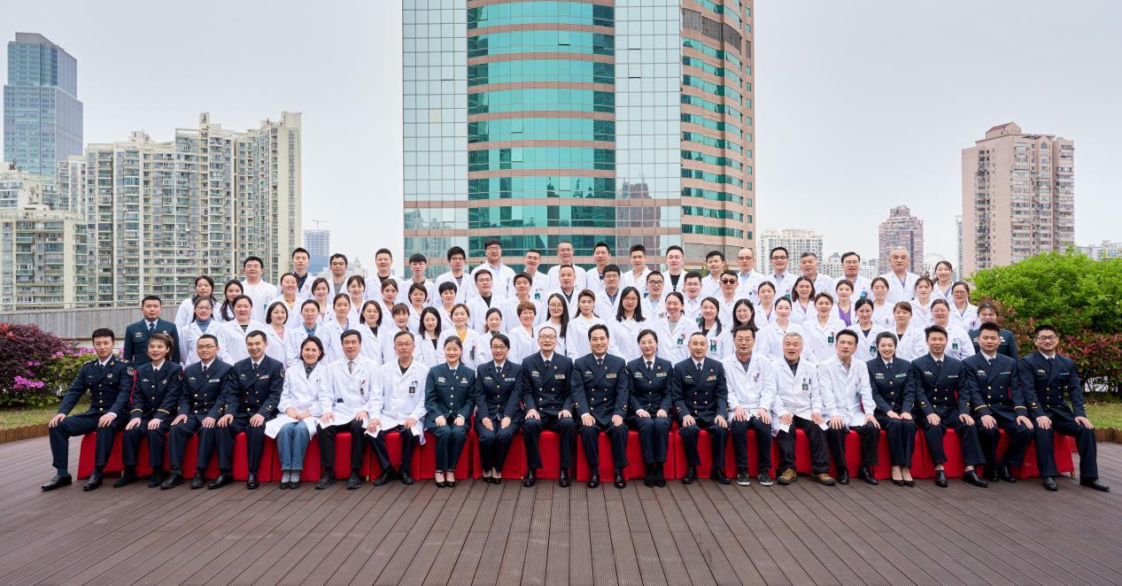 Group photo of shanghai changzheng hospital Radiological Diagnosis Department
Over the years, under the leadership of Professor Liu Shiyuan, the Department of Radiological Diagnosis in shanghai changzheng hospital has become a characteristic discipline with complete sub-specialties, strong technical force, reasonable echelon structure and balanced development of medical teaching and research, and plays an important role in the field of image diagnosis in China. Nowadays, the department has become the key discipline of the Ministry of Education and the lung cancer center of the whole army. In the comprehensive ranking list of China Hospital in Fudan Edition in 2019, this department ranked 11th.
"The rapid development of medical imaging has enabled patients to have a better sense of acquisition, and our diagnostic ability and treatment level have achieved a high-level leap." Looking to the future, Professor Liu Shiyuan has a bigger plan: focusing on the imaging diagnosis of chest diseases and the clinical application of artificial intelligence, expanding the research and development of new technologies for neurological diseases and bone and joint imaging, insisting on the synergy of clinical and scientific research, innovating and developing, and constantly achieving comprehensive breakthroughs in medical treatment, teaching, scientific research and combat readiness.
